File:Chest high-resolution computed tomography.jpg
No higher resolution available.
Chest_high-resolution_computed_tomography.jpg (783 × 493 pixels, file size: 99 KB, MIME type: image/jpeg)
Summary
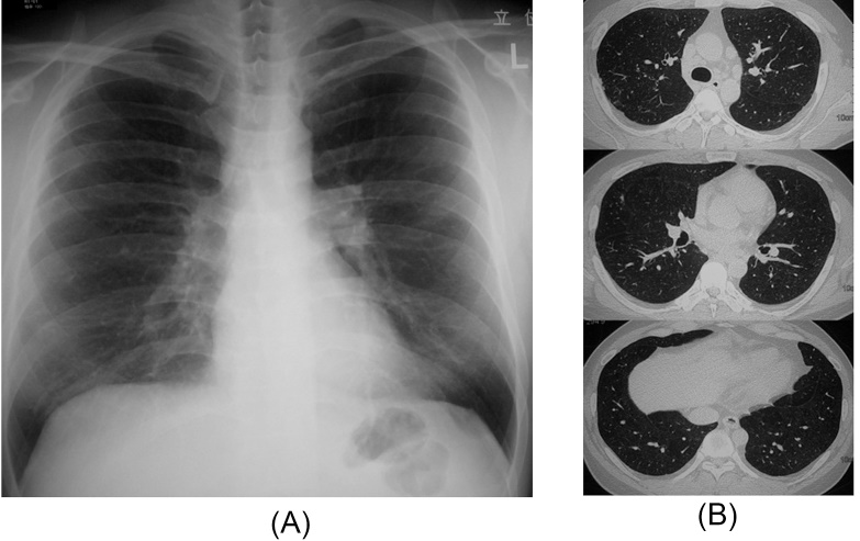
| Description | English: A) Chest X-ray showed faint nodular shadows in the bilateral lung fields. (B) Chest high-resolution computed tomography shows diffuse fine nodular opacities that were distributed mainly in the upper and middle lung zones with mediastinal lymph node enlargement. |
| Date | 16 March 2017 () |
| File source | https://www.ncbi.nlm.nih.gov/pmc/articles/PMC4901174/ |
| Author | Hiromi Tomioka, Toshihiko Kaneda, Eiji Katsuyama Masanori Kitaichi, Hiroshi Moriyama, and Eiichi Suzukie |
Licensing
|
|
File history
Click on a date/time to view the file as it appeared at that time.
| Date/Time | Thumbnail | Dimensions | User | Comment | |
|---|---|---|---|---|---|
| current | 00:30, 17 March 2017 | | 783 × 493 (99 KB) | SaminderjitNagi (talk | contribs) | |
| 00:25, 17 March 2017 |  | 256 × 256 (5 KB) | SaminderjitNagi (talk | contribs) | User created page with UploadWizard |
You cannot overwrite this file.
File usage
The following page uses this file: