Carry, a 29-year-old woman, is 32 weeks pregnant with her first child. As a foodie, she loves trying different gourmet restaurants in Vancouver. A new “farm-to-table” restaurant has opened and she gets a bunch of friends to go out for a nice dinner. Carry orders a toasted pecan, strawberry and mature goat cheese salad to start. She knows that, in pregnancy, she should not eat raw or unpasteurized cheeses but cannot help the temptation - she’s heard that this is the best salad in Vancouver. A few days later, she develops mild diarrhea and night sweats that she thinks will eventually pass, but the following day she has a fever so she goes to the emergency department where she has blood and stool cultures collected. The blood cultures turn positive for Listeria monocytogenes. She wonders what effect this will have on her unborn baby.
The Body System
(i) Describe the signs (objective characteristics usually detected by a healthcare professional) and symptoms (subjective characteristics experienced by the patient). Are there any other signs or symptoms that could have been commented on but are not presented in the case? What are the key History of Presenting Illness elements presented? What laboratory samples are taken and why?
Listeria monocytogenes, which is the cause of Listeriosis, is a Gram-positive, facultatively anaerobic, opportunistic, and life-threatening food-borne illness particularly found in dairy and leafy green products [1, 2]. Listeria causes invasive or intestinal illness [3]. Invasive illness occurs Listeria disseminates out of the intestines [3]. Symptoms usually arise within 2 weeks of consumption of contaminated food [3] but can begin between 3 to 70 days post-exposure [4]. Symptoms of invasive illness in pregnant people include fever and flu-like symptoms including muscle aches and fatigue [3]. Invasive illness in non-pregnant people may lead to the aforementioned symptoms as well as headache, stiff neck, confusion, loss of balance, and seizures [3]. Symptoms of invasive tend to be asymptomatic or mild in pregnant women and severe in non-pregnant individuals [3]. Symptoms of the intestinal illness include mild diarrhea and vomiting [3].
Depending on the host and consumption of Listeria, incubation times can vary; generally, gastroenteritis appears within 24 hours while the appearance of invasive disease can take around 11 days [5]. Incubation periods tend to be longer in pregnant people versus non-pregnant people [5]. Febrile gastroenteritis secondary to Listeria infection can occur in both healthy and immunocompromised patients [5]. For this manifestation to occur, there needs to be a large amount of Listeria ingested from implicated foods [5]. Symptoms include fever, nausea, cramps, diarrhea, vomiting, headache, constipation, and muscle aches [1]. Febrile gastroenteritis is not considered an invasive disease and clears spontaneously within 1 to 3 days [5]. Severe cases of Listeriosis involve the bacterium spreading to the nervous system causing meningitis, which includes symptoms such as a stiff neck, confusion, headache, and loss of balance. [1,2]. Additionally, symptoms may vary with the infected person. For example, if the infection is transmitted to the fetus during pregnancy the bacterium can be detected from the amniotic fluid, amnionitis [6]. An L. monocytogenes infection can spread into the bloodstream, becoming septic [6].
In this case, Carry’s signs and symptoms are able to be grouped together; she experiences diarrhea, night sweats, and fever. Symptoms such as nausea, muscle aches, and headaches are not mentioned. Additionally, there is no comment on symptoms relating to her pregnancy. Symptoms of invasive illness in pregnant people include fever and flu-like symptoms including muscle aches and fatigue [3]; however, an infection can negatively impact the pregnancy: miscarriage, stillbirth, premature delivery, or life-threatening infection of the infant [2]. Since Carry is pregnant and experienced diarrhea and fever, she likely has invasive Listeria.
A few key histories of presenting illness elements are presented in the case study: a few days after eating goat cheese at a Vancouver restaurant, Carry develops mild diarrhea and night sweats. However, the next day she has a fever and goes to the hospital. Samples are collected and her treatment plan will be solidified after diagnosis.
The key components of a history of presenting illness are location, quality, severity, duration, timing, context, modifying factors, and associated signs and symptoms [7]. For this case, the key history of presenting illness elements are as follows:
Location: There is no location of pain or discomfort explicitly reported. However, Carry’s reported experience of diarrhea implies the involvement of the gastrointestinal tract is affected, while fever and night sweats suggest a systemic body reaction.
Quality: The quality of Carry’s symptoms are reported to be mild. The fact that her night sweats developed into a fever could possibly indicate that her symptoms are progressing from mild to severe. It is also noted that pregnant women tend to only experience fever and other nonspecific symptoms [8].
Severity: The severity of Carry’s symptoms is reported to be mild but may be on the path toward being more severe.
Timing: Carry’s symptoms developed a few days after she consumed goat cheese. Her diarrhea and night sweats presented first, while her fever began one day after symptom onset. This timing lines up with the typical Legionella infection which occurs within a few days to a few weeks after infection [7].
Context: Carry was likely infected upon eating her which contained raw or unpasteurized cheeses and leafy greens. All surface-ripened goat cheeses are made from raw, unpasteurized goat milk [9]. Consumption of raw unpasteurized cheeses is not recommended in pregnancy as it carries between a 50- to 160-fold higher risk for contamination with Listeria monocytogenes (L. monocytogenes) [10, 11].
Modifying Factors: There is no mention of Carry’s previous medical history in the case, so it is safe to assume she is not immunosuppressed, however, she is pregnant which puts her at more of a risk for infection [8]. General treatment to help the infection revolves around antibiotics such as penicillin and ampicillin, although studies have shown that there is a rise in antibiotic-resistant strains of the bacteria [12].
Associated manifestations: based on the case we can not tell if Carry has any associated manifestations until further testing is done. Some associated manifestations include meningitis and rhombencephalitis which are both uncommon and often appear in immunocompromised patients [13].
Diagnosis of L. monocytogenes infection depends on the laboratory identification of the bacterium from blood or cerebrospinal fluid [13]. Blood cultures are taken to detect the bacteria in the bloodstream, which can help diagnose invasive listeriosis [3,4]. It is common for pregnant women to have this form of listeriosis [3]. Stool cultures can help identify the bacteria in the feces, which may suggest a gastrointestinal form of the infection, though the diagnosis of invasive Listeria infection is not made based on stool samples, so it is important to take other body fluid samples for culture [3, 14]. It is important to test for these differences because they can cause different symptoms and thus require different types of t treatment. Utilizing cerebrospinal fluid is only useful if the infection has spread to the nervous system like in rhombencephalitis syndrome [13]. However, a Gram stain of the sample reveals the characteristic Gram-positive coccobacilli, allowing for accurate identification and diagnosis [13]. However, in cases where antibiotic therapy is occurring, laboratory samples are taken from nonsterile sites like stool or vaginal culture [13].
References:
Canada PHAof. 2016. Government of Canada. Canadaca. / Gouvernement du Canada. Accessed on March 29, 2023 from https://www.canada.ca/en/public-health/services/diseases/listeriosis/symptoms-listeriosis.html
Centers for Disease Control and Prevention. 2012. Clinical features/signs and symptoms. Centers for Disease Control and Prevention. Centers for Disease Control and Prevention. Accessed on March 29, 2023 from https://www.cdc.gov/listeria/outbreaks/cheese-09-12/signs-symptoms.html#:~:text=The%20symptoms%20vary%20with%20the, and% 20 other%20non%2Dspecific%20symptoms.
CDC Centers for Disease Control and Prevention. Listeria (Listeriosis): Symptoms [Internet]. CDC Centers for Disease Control and Prevention; 2022 May 3. Available from: https://www.cdc.gov/listeria/symptoms.html
Canada.ca. Symptoms of listeriosis (Listeria) [Internet]. Government of Canada; 2016 Aug 10. Available from: https://www.canada.ca/en/public-health/services/diseases/listeriosis/symptoms-listeriosis.html
Collins JP, Griffin PM. Listeria monocytogenes Infections. In: Loscalzo J, Fauci A, Kasper D, Hauser S, Longo D, Jameson J. eds. Harrison's Principles of Internal Medicine, 21e. McGraw Hill; 2022. Accessed March 30, 2023. https://accessmedicine.mhmedical.com/content.aspx?bookid=3095§ionid=263549003
Osmosis. Congenital torch infections: Pathology review | osmosis. Accessed on March 29, 2023 from https://www.osmosis.org/learn/Congenital_TORCH_infections:_Pathology_review#:~:text=The%20next%20TORCH%20infections%20are,varicella%20zoster%20virus%2C%20and%20listeria.
Chan L, Lin H, Hsiao S. Successful treatment of maternal listeria monocytogenes bacteremia in the first trimester of pregnancy: A case report and literature review. Taiwanese journal of obstetrics & gynecology. 2018;57:462-463.
Committee Opinion No. 614: Management of Pregnant Women With Presumptive Exposure to Listeria monocytogenes. Obstetrics and gynecology (New York. 1953). 2014;124:1241-1244.
Healthline. Is Goat Cheese Safe During Pregnancy? [Internet]. New York, NY: Healthline Media; 2020 May 29. https://www.healthline.com/nutrition/goat-cheese-pregnancy#types-to-avoid
CDC Centers for Disease Control and Prevention. Listeria (Listeriosis): Prevent Listeria [Internet]. CDC Centers for Disease Control and Prevention; 2023 Mar 15. Available from: https://www.cdc.gov/listeria/prevention.html
Jackson K, Gould L, Hunter JC, et al. Listeriosis Outbreaks Associated with Soft Cheeses, United States, 1998–2014. Emerg. Infect. Dis. 2018;24(6):1116-1118. doi:10.3201/eid2406.171051.
Lungu B, O'Bryan CA, Muthaiyan A, et al. Listeria monocytogenes: Antibiotic Resistance in Food Production. Foodborne pathogens and disease. 2011;8:569-578.
Schlech WF. 2019. Epidemiology and clinical manifestations of listeria monocytogenes infection. Microbiology Spectrum 7. https://doi.org/10.1128/microbiolspec.GPP3-0014-2018
Diagnosis and treatment. Centers for Disease Control and Prevention. https://www.cdc.gov/listeria/diagnosis.html. Published May 3, 2022. Accessed March 30, 2023.
(ii) What are the two major clinical syndromes of listeria and which body systems can be affected? In what way has the normal physiological functioning of this body system been disturbed by the infection (specifically looking at the physiological changes without detailing the bacterial mechanism of this disturbance as that is the basis of another question). Representing this diagrammatically is helpful to demonstrate understanding.
The two major clinical syndromes of Listeria infection are non-invasive intestinal illness and invasive illness [1]. Non-invasive Listeria leads to febrile gastroenteritis [2]. L. monocytogenes first infects intestinal epithelial cells [3]. L. monocytogenes uses M cells of the intestine and intestinal villi cells as a point of entry [4]. Basolateral spread from infected intestinal cells to enterocytes causes enteritis and leads to gross intestinal lesions [4]. A diagram of the progression from Listeria introduction to the host to invasive disease can be seen in Figure 1 [C].
Figure 1. Introduction of Listeria monocytogenes to the host and its progression to invasive infection. [5]
L. monocytogenes is multisystemic and can infect various tissues [3]. After infection with Listeria monocytogenes, the bacteria move from the gut to the mesenteric lymph nodes, targeting the liver and spleen [C]. From there, if the immune response is unable to control the infection, the bacteria move into the bloodstream and can infect host organs, or the secondary target organs [C]. L. monocytogenes also infect macrophages [5]. After phagocytosis, L. monocytogenes lyses the phagosome [6]. In the cell, L. monocytogenes disrupt normal processes by modulating actin polymerization and initiates a cell-mediated immune response [6]. L. monocytogenes is translocated from the intestine to deeper organs quickly [4]. Invasive illness results from the disseminated or systemic spread of Listeria and may lead to various syndromes including meningitis, meningoencephalitis, encephalitis, rhombencephalitis, sepsis, endocarditis, peritonitis, myocarditis, pneumonia, pleuritis, sinusitis, conjunctivitis, ophthalmitis, septic arthritis, biliary tract disease, septicemia, hepatitis, liver abscess, endophthalmitis, febrile gastroenteritis, and osteomyelitis [3]. These impact the brain, heart, abdomen, lungs, sinuses, eyes, joints, gallbladder, bone, liver, and intestine.
Figure 2. Mid-sagittal view of the brain showing the meninges. Bacteria are shown being able to reach the meninges through the blood–CSF barrier which are in close anatomical relation to the cerebral cortex and brain parenchyma.[7]
In the liver, neutrophil recruitment to infected cells causes microabscess formation and destruction of infected hepatocytes [4]. In pregnant women, Listeria penetrates the placenta causing inflammatory infiltration, necrosis, microabscesses, and focal necrotizing villitis [4]. Infection may spread to the fetus, causing fetal death or premature birth and military pyogranulomatous lesions [4]. In the brain, Listeria causes meningitis with inflammatory infiltrates and infectious foci in the brain parenchyma, which may lead to focal necrosis, microabscesses, and macroscopic brain lesions [4]. Brain lesions may cause unilateral cranial nerve paralysis [4].
Figure 3. Pathophysiology and associated mechanisms of bacterial meningitis shown along with its various pathways of infection. [9]
Meningitis is an infection of the meninges, the protective layers that surround the brain and spinal cord that primarily occurs in neonates and older adults [7]. Neonates acquire early-onset infection in utero or late-onset within a few weeks of birth [8]. Adults acquire the infection through eating contaminated food. For a bloodborne pathogen, such as listeria, the pathway of invasion is assumed to be through the subarachnoid space and is a multi-step process that eventually results in the traversal of the blood-brain barrier [7]. The mechanism of Listeria meningitis is as follows: the bacterium enters the host epithelial cells and hepatocytes [8]. After phagocytosis, the bacterium activates rapid lysis of the phagolysosome, releasing the Listeria into the cytoplasm [8]. There the organism replicates and invades adjacent cells. Eventually, the bacterium crosses the meninges and blood-brain barrier, allowing the organism to grow within the brain [8]. Within the brain, Listeria bacteria infect the brain tissue, which classifies this syndrome as meningoencephalitis and can be found in the brain parenchyma, the blood vessels, and the meninges [C]. Listeria meningitis affects the central nervous system by causing excessive inflammation of the meninges, a protective membrane covering the brain parenchyma and spinal cord [9]. This excessive inflammation increases the pressure and volume of the cerebrospinal fluid, impeding neural functions. Common symptoms of L. monocytogenes meningitis include headache, fever, stiff neck, sensitivity to light, and in severe cases, seizures, alteration of consciousness, and brain abscess [7,9]. Morbidity of Listeria-associated meningoencephalitis is associated with ventriculitis and small periventricular abscesses, as well as ventricular inflammation [C]. Figures 2 and 3 show the pathways for meningitis infection as well as its pathophysiology in detail.
L. monocytogenes rhombencephalitis is characterized by progressive brainstem dysfunction [9]. Symptoms appear within four to ten days of infection, headache, muscle aches, nausea, and vomiting [9]. Afterward, defects of the cranial nerve and hemiparesis occur impacting cerebellar signaling and sensory functions [9]. Typical Listeria rhombencephalitis occurs due to the proliferation and spread of L. monocytogenes in the intracerebral tracts [11]. Infection can also result in lesions in the brainstem and cerebellum [11]. Figure 4 shows the increased enhancement of arachnoid matter (white arrows) which appear to be inflamed as a result of Rhombencephalitis.
References:
Figure 4. Sagittal plane MRI of arachnoid mater (white arrows). Image after contrast injection demonstrated leptomeningeal enhancement which was indicating inflammation. [10]CDC Centers for Disease Control and Prevention. Listeria (Listeriosis): Symptoms [Internet]. CDC Centers for Disease Control and Prevention; 2022 May 3. Available from: https://www.cdc.gov/listeria/symptoms.html
World Health Organization. Listeriosis [Internet]. World Health Organization; 2018 Feb 20. Available from: https://www.who.int/news-room/fact-sheets/detail/listeriosis
Krawczyk-Balska A, Markiewicz Z. The intrinsic cephalosporin resistome of Listeria monocytogenes in the context of stress response, gene regulation, pathogenesis and therapeutics. J Appl Microbiol. 2016 Feb;120(2):251-65. doi: 10.1111/jam.12989.
Vázquez-Boland JA, Kuhn M, Berche P, Chakraborty T, Domínguez-Bernal G, Goebel W, et al. Listeria pathogenesis and molecular virulence determinants. Clin Microbiol Rev. 2001 Jul;14(3):584-640. doi: 10.1128/CMR.14.3.584-640.2001.
Schlech WF. Epidemiology and Clinical Manifestations of Listeria monocytogenes Infection. Microbiol Spectr. 2019;7(3). doi: 10.1128/microbiolspec.GPP3-0014-2018.
Van de Beek, D., Brouwer, M., Hasbun, R. et al. Community-acquired bacterial meningitis. Nat Rev Dis Primers 2, 16074 (2016). https://doi.org/10.1038/nrdp.2016.74
Tessier JM, Scheld WM. 2015. Bacterial infections of the central nervous system. Molecular Medical Microbiology 1693–1707
Disson O, Lecuit M. 2012. Targeting of the central nervous system bylisteria monocytogenes. Virulence 3:213–221.
Jubelt B, Mihai C, Li TM, Veerapaneni P. Rhombencephalitis / Brainstem Encephalitis. Current neurology and neuroscience reports. 2011;11:543-552.
Ashraf VV, Salam KA. 2021. Listeria rhombencephalitis. Journal of Neurosciences in Rural Practice 12:443–444.
(iii) What antibiotics/treatments might have been given (i.e., what are antibacterial treatments and how do these treatments work to help the body clear the organism)? Why do cephalosporins not work for Listeria? Representing this diagrammatically is helpful to demonstrate understanding.
Patients with invasive Listeria are usually treated with antibiotics [1]. Intestinal illness does not usually require antibiotics and instead involves hydration with adequate fluids to prevent dehydration [1]. In intestinal illness, antibiotics are used for patients who are very ill or at risk of becoming very ill [1]. Early treatment of pregnant mothers prevents transplacental infection of the fetus [2]. Commonly used antibiotics for Listeria monocytogenes infections include ampicillin and penicillin G. Most β-lactam antibiotics, except cephalosporins, are effective against L. monocytogenes [2].These antibiotics are effective against Listeria because they target the bacterial cell wall or protein synthesis, which are essential for the bacteria's survival and reproduction [3]. By inhibiting the enzymes responsible for cross-linking the peptidoglycan chains that form the cell wall, the cell wall weakens, causing the bacterial cell to rupture and die [3]. Another viable option is trimethoprim-sulfamethoxazole (TMP-SMX). TMP-SMX is a combination of two antibiotics that work by inhibiting bacterial metabolism [4]. Sulfamethoxazole blocks the synthesis of folic acid, a vitamin essential for bacterial growth and reproduction, while trimethoprim blocks the enzyme that converts dihydrofolic acid to tetrahydrofolic acid, a necessary cofactor for the production of DNA, RNA, and proteins [4].
Most Listeria antibiotics are bacteriostatic, temporarily suppress bacterial growth, while few are bactericidal [5]. A combination treatment of ampicillin and gentamicin is the current standard therapy [2, 5]. A Listeria infection persisting with Meningitis recommends a drug regimen of intravenous ampicillin or penicillin with gentamicin for three weeks [6,7]. Patients allergic to penicillin are prescribed trimethoprim-sulfamethoxazole [6]. A Listeria infection persisting without meningitis utilizes a drug regimen of intravenous ampicillin or penicillin with gentamicin for two weeks [7]. However, in the case of a brain abscess with rhombencephalitis or cerebritis, the drug regimen is recommended for four to six weeks [7].
Ampicillin is the drug of choice for a Listeria infection. Ampicillin is bacteriostatic and amplifies the gentamicin’s bactericidal effects [5]. Ampicillin is a synthetic penicillin derivative that inhibits bacterial cell wall synthesis to induce cell lysis [8]. Ampicillin is dosed at least 6g IV per day in divided doses [D]. The mechanism of action for ampicillin is to bind to penicillin-binding proteins (PBPs) located in the bacterial cell wall [8]. Afterward, ampicillin inhibits bacterial cell wall synthesis, by interfering with the cross-linkage of peptidoglycan chains [9]. This inhibition interferes with autolysin enzyme activity, resulting in cell lysis [8, 9]. However, recent research indicates ampicillin treatment may intrinsically promote drug-resistant forms of L. monocytogenes [10].
Gentamicin is an aminoglycoside that targets Listeria’s 30S ribosome to hinder protein translation, which disrupts cell membrane integrity [11]. The addition of gentamicin is to synergize with ampicillin and amplifies its effects [7]. Gentamicin activates macrophages to target the listeria bacterium [12]. Therefore, significantly increasing the antibiotic effect on inhibiting the spread of infection. The synergism from combining ampicillin and gentamicin is primarily utilized in the treatment of high-risk patients like newborns or immunocompromised individuals [13]. Gentamycin is not always a favorable addition because it can cause severe renal toxicity. Recent research indicates mutations in the atpG2 – a gene involved in the ATP synthase construct – may promote resistance to gentamicin in L. monocytogenes [14,D].
L. monocytogenes exhibits intrinsic resistance to cephalosporin antibiotics encoded by a bacterial resistome; it does not result from antibiotic selective pressure, horizontal gene transfer, or mutation [5]. This is problematic as cephalosporins are often used for idiopathic sepsis [5]. The intrinsic resistome encodes multidrug resistance efflux pumps, which pump the antibiotic out of the cell, proteins involved in peptidoglycan synthesis and modification, detoxifying cell envelope proteins such as AnrB, TelA, OatA, Lmo2522, and Lmo1941, cytoplasmic proteins Lmo1416, Lmo2210, Lmo2442 and Lmo2568 of unknown function, and regulatory proteins [5]. The main cause of L. monocytogenes resistance to cephalosporins is that it lacks penicillin-binding proteins (PBP) that can be bound and inhibited by cephalosporin [5]. Normally, cephalosporins bind to PBP to inhibit its transpeptidase and carboxypeptidase functionalities, leading to a loss of peptidoglycan integrity and cell death [5]. Cephalosporins, while sharing a similar mechanism of action with penicillins such as ampicillin, are inactive against Listeria monocytogenes due to ineffective binding to PBP3 [E]. In addition to these factors, cephalosporins do not cover gram-positive bacteria, which is the group that Listeria belongs to. Cephalosporins are designed to target gram-negative bacteria, which have a different cell wall structure compared to gram-positive bacteria [5].
References:
CDC Centers for Disease Control and Prevention. Listeria (Listeriosis): Diagnosis and Treatment [Internet]. CDC Centers for Disease Control and Prevention; 2022 May 3. Available from: https://www.cdc.gov/listeria/diagnosis.html
Schlech WF. Epidemiology and Clinical Manifestations of Listeria monocytogenes Infection. Microbiol Spectr. 2019;7(3). doi: 10.1128/microbiolspec.GPP3-0014-2018.
Rodríguez-Villodres Á, Lepe JA, Blázquez J, Aznar J. Effect of subinhibitory concentrations of ampicillin on Listeria monocytogenes. Enfermedades infecciosas y microbiologia clinica (English ed.). 2020;38:72-75.
Stavropoulos C, Tolentino B, Woods K, Pyburn D, Patterson S, Jean R. Listeria Rhomboencephalitis in an Immunocompetent Host: Treatment With Trimethoprim-Sulfamethoxazole and Ampicillin: A Case Report and Review of Treatment Options. Infectious diseases in clinical practice (Baltimore, Md.). 2021;29:e204-e207.
Krawczyk-Balska A, Markiewicz Z. The intrinsic cephalosporin resistome of Listeria monocytogenes in the context of stress response, gene regulation, pathogenesis and therapeutics. J Appl Microbiol. 2016 Feb;120(2):251-65. doi: 10.1111/jam.12989.
Rogalla D, Bomar PA. Listeria Monocytogenes. 2022. In: StatPearls [Internet]. Treasure Island (FL): StatPearls Publishing. Accessed on March 29, 2023 from https://www.ncbi.nlm.nih.gov/books/NBK534838/
Shoham S. n.d. Listeria monocytogenes: Johns Hopkins Abx Guide. Listeria Monocytogenes | Johns Hopkins ABX Guide. Accessed on March 29, 2023 from https://www.hopkinsguides.com/hopkins/view/Johns_Hopkins_ABX_Guide/540318/all/Listeria_Monocytogenes
DrugBank. Ampicillin [Internet]. DrugBank; [cited 2023 Mar 29]. Available from: https://go.drugbank.com/drugs/DB00415
PubChem. n.d. Ampicillin. National Center for Biotechnology Information PubChem Compound Database. U.S. National Library of Medicine. Accessed on March 30, 2023 from https://pubchem.ncbi.nlm.nih.gov/compound/Ampicillin#section=Names-and-Identifiers
Grosboillot V, Keller I, Ernst C, Loessner MJ, Schuppler M. 2022. Ampicillin treatment of intracellular listeria monocytogenes triggers formation of persistent, drug-resistant L-form cells. Frontiers in Cellular and Infection Microbiology 12.
DrugBank. Gentamicin [Internet]. DrugBank; [cited 2023 Mar 29]. Available from: https://go.drugbank.com/drugs/DB00798
Drevets DA, Canono BP, Leenen PJ, Campbell PA. 1994. Gentamicin kills intracellular listeria monocytogenes. Infection and Immunity 62:2222–2228.
Azimi PH, Koranyi K, Lindsey KD. 1979. listeria monocytogenes: Synergistic effects of Ampicillin and gentamicin. American Journal of Clinical Pathology 72:974–977.
Ng JM, Ngeow YF, Saw SH, Ng HF, Zin T. 2022. Mutations in ATPG2 may confer resistance to gentamicin in listeria monocytogenes. Journal of Medical Microbiology 71.
(iv) What are ToRCH/SCoRCH infections and why are they important? Why is Listeria not a ToRCH/SCoRCH infection? Why are pregnant women at higher risk of listeria? Other than pregnant people, what are other high risk groups for invasive disease? “How should one prevent exposure to listeria?”
ToRCH/SCoRCH refers to a group of diseases that causes congenital conditions if a fetus is exposed to them in utero. These infections are passed vertically from an infected mother to a developing neonate through the placenta or perinatally during or after birth [1]. ToRCH stands for toxoplasmosis, other (including Treponema pallidum, varicella, human immunodeficiency virus, parvovirus B19, and ZIKV), rubella, cytomegalovirus, and herpes simplex virus [2, 3]. ToRCH diseases include syphilis, cytomegalovirus (CMV), ‘other’, rubella, toxoplasmosis, chickenpox, herpes simplex virus (HSV), and blood-borne viruses [4]. A SCoRCH diagnostic approach considers signs of congenital infection, serological testing details, and direct diagnostic methods [4]. SCoRCH is important as it assists with the diagnosis of perinatal infections that can lead to miscarriage, stillbirth, or congenital abnormalities in the developing fetus [3].
Pregnant women have a 10-fold higher risk of Listeria infection [5], and 13 to 100 times greater risk of invasive infection [6]. Severe, life-threatening infection is rare in pregnant women [6]. Listeria poses a risk to the neonate; it can cause miscarriage, stillbirth, preterm birth, or serious neonatal disease [6]. L. monocytogenes neonatal infection is not directly associated with any specific congenital birth defects, unlike other ToRCH pathogens [7], so Listeria is not a ToRCH infection. Listeria is not considered a SCoRCH infection as fetal complications are regarded as secondary to the mother’s illness, which can be severe and life-threatening without prompt diagnosis and treatment [8]. Additionally, it is hypothesized that any invasive bacteria that can survive in host cells can colonize the placenta and fetus [8]. Therefore, L. monocytogenes is recognized as a pathogen that can cross the placenta but is not part of the SCoRCH group [9].
Listeria is not a ToRCH/SCoRCH infection because it is not typically transmitted vertically from mother to fetus, and it is not typically associated with congenital abnormalities in the fetus. Pregnant women are at higher risk of listeria because their immune systems are suppressed during pregnancy, making them more vulnerable to infections; these changes are thought to be related to the body's need to tolerate the developing fetus, which is partially comprised of foreign DNA [1, 2]. Additionally, pregnancy is associated with changes in the gastrointestinal tract that can make it easier for Listeria to colonize the gut and potentially cause infection, which would explain Carry’s symptoms [1].
Other high-risk groups include adults aged 65 years or older, newborns of infected mothers, and immunocompromised people [10, 11]. Invasive listeriosis affects high-risk groups such as pregnant women, older adults, infants, and patients undergoing treatment for cancer, AIDS, and organ transplants [12, 13]. Adults over the age of 65 are 4.4% more likely to be infected than the general population [12]. The mortality rate of high-risk groups with Listeria infection is 20%-30% [13]. Increased susceptibility is largely due to immune suppression; immunosuppressive agents predispose individuals to invasive infection [14]. Hormonal fluctuations in pregnancy modulate immunity. Estradiol impacts innate, cell-mediated, and humoral immunity and progesterone suppresses the immune response [6]. Increasing levels of estrogen and progesterone lead to thymic involution, leading to diminished T cell responses, accompanied by a shift from Th1 to Th2 responses [6]. Control of Listeria infection is largely dependent on effective macrophages [14]. Th1 cells activate macrophages [12] but are present in lower numbers as pregnancy favors Th2 cells [6]. Pregnant women exhibit both an impaired cell-mediated immune response and decreased gastrointestinal motility, which contributes to a higher risk of invasive Listeria infection [14]. Additionally, L. monocytogenes exhibit uterus and placenta epithelial tropism, hence the occurrence of infection increases during pregnancy [10].
Listeria monocytogenes are transmitted to human hosts through contaminated foods [15]. To prevent exposure to Listeria the CDC recommends that people who may have compromised immune systems, such as the elderly, those with immunocompromising conditions or drugs, and pregnant people avoid certain foods such as unpasteurized soft cheese, unheated deli meat, premade salads, refrigerated meat spreads and smoked fish, raw sprouts, certain melons, and unpasteurized milk products [15]. A comprehensive table can be seen in Figure 1 [15]. These foods are more likely to be contaminated with Listeria monocytogenes either due to ideal growing conditions for the bacteria or a higher likelihood of contamination [15]. Another method to prevent exposure to Listeria is to ensure foods are cooked thoroughly and properly; actions such as cooking, fermenting, and drying kill Listeria monocytogenes bacteria. Although refrigeration is not protective against colonized bacteria, it can help to prevent Listeria colonization of foods [15]. One should ensure that refrigerators are kept at or below 40°F (4°C) and freezers should be kept at 0°F (-18°C) to prevent L. monocytogenes growth, and keep the refrigerator clean so that there are no areas for Listeria to grow and spread [16]. To reduce one’s risk of disease, one should follow general food safety and hygiene tips and wash their hands frequently [17].
Food processing plants also take measures to try and prevent Listeria monocytogenes contamination in their products. Benzalkonium chloride and chlorine-based biocides are often used to sterilize areas where there is direct food contact, though studies show that continuous use with these products is leading to tolerance from Listeria to these products [18]. This is due to the frequent application of less than adequate amounts of the biocides [18]. Solutions to this problem that have been proposed are alternating between biocides with different mechanisms of action, in order to prevent resistance from forming, as well as ensuring that there is an adequate biocide used to disinfect the area [18]. These ideals can be extrapolated to use in delis as well, as even food that has been heated thoroughly to kill Listeria bacteria can be recontaminated if reintroduced to a contaminated surface or utensil [15].
If one suspects that they got food poisoning from a restaurant, or if they notice unclean restaurants or grocery stores, they should contact their local public health authority [17]. This may prevent others from getting sick.
References
Leeper C, Lutzkanin A 3rd. Infections During Pregnancy. Prim Care. 2018 Sep;45(3):567-586. doi: 10.1016/j.pop.2018.05.013.
Coyne, C., Lazear, H. Zika virus — reigniting the TORCH. Nat Rev Microbiol. 2016 Aug 30;14:707–715. https://doi.org/10.1038/nrmicro.2016.125
Neu N, Duchon J, Zachariah P. TORCH infections. Clin Perinatol. 2015 Mar;42(1):77-103, viii. doi: 10.1016/j.clp.2014.11.001.
Penner J, Hernstadt H, Burns JE, Randell P, Lyall H. 2020. Stop, think scortch: Rethinking the traditional ‘torch’ screen in an era of re-emerging syphilis. Archives of Disease in Childhood 106:117–124.
CDC Centers for Disease Control and Prevention. Listeria (Listeriosis): People at Risk – Pregnant Women and Newborns [Internet]. CDC Centers for Disease Control and Prevention; 2022 Oct 25. Available from: https://www.cdc.gov/listeria/risk-groups/pregnant-women.html
Kourtis AP, Read JS, Jamieson DJ. Pregnancy and Infection. The New England journal of medicine. 2014;370:2211-2218.
Belanger BG, Lui F. Embryology, Teratology TORCH. [Updated 2022 Jul 25]. In: StatPearls [Internet]. Treasure Island (FL): StatPearls Publishing; 2023 Jan-. Available from: https://www.ncbi.nlm.nih.gov/books/NBK545148/
Vázquez-Boland JA, Krypotou E, Scortti M. 2017. listeria placental infection. mBio
MedBullets. n.d. Torches infections. ToRCHeS Infections - Microbiology - Medbullets Step 1. Accessed on March 29, 2023 from https://step1.medbullets.com/microbiology/104120/torches-infections
Jackson K, Gould L, Hunter JC, et al. Listeriosis Outbreaks Associated with Soft Cheeses, United States, 1998–2014. Emerg. Infect. Dis. 2018;24(6):1116-1118. doi:10.3201/eid2406.171051.
CDC Centers for Disease Control and Prevention. Listeria (Listeriosis): People at Risk [Internet]. CDC Centers for Disease Control and Prevention; 2022 Nov 8. Available from: https://www.cdc.gov/listeria/risk.html
Centers for Disease Control and Prevention (CDC)2013. Vital signs: listeria illnesses, deaths, and outbreaks—United States, 2009-2011. Annals of Emergency Medicine 62:536–537.
World Health Organization. Listeriosis. World Health Organization. World Health Organization. Accessed on March 29, 2023 from https://www.who.int/news-room/fact-sheets/detail/listeriosis#:~:text=doses%20of%20L.-,monocytogenes.,transplants%2C%20elderly%20people%20and%20infants.
Schlech WF. Epidemiology and Clinical Manifestations of Listeria monocytogenes Infection. Microbiol Spectr. 2019;7(3). doi: 10.1128/microbiolspec.GPP3-0014-2018.
CDC Centers for Disease Control and Prevention. Listeria (Listeriosis): Prevent Listeria [Internet]. CDC Centers for Disease Control and Prevention; 2023 Mar 15. Available from: https://www.cdc.gov/listeria/prevention.html
FDA. What you need to know about preventing Listeria infections [Internet]. U.S. Food & Drug Administration; 2018 Mar 22. Available from: https://www.fda.gov/food/buy-store-serve-safe-food/what-you-need-know-about-preventing-listeria-infections
Canada.ca. Prevention of listeriosis (Listeria) [Internet]. Public Health Agency of Canada, Government of Canada; 2016 Aug 10. Available from: https://www.canada.ca/en/public-health/services/diseases/listeriosis/prevention-listeriosis.html
Duze ST, Marimani M, Patel M. Tolerance of Listeria monocytogenes to biocides used in food processing environments. Food Microbiol. 2021;97:103758. doi:10.1016/j.fm.2021.103758
The Microbiology Laboratory
(i) What are the most common bacterial pathogens associated with fever and diarrhea?
The most common bacterial pathogens that are associated with both diarrhea and fevers are: E. coli, Shigella, Salmonella, Campylobacter, Yersinia, Listeria, Clostridium spp., and Vibrio cholerae (1). All of these listed bacteria either infect the large or small intestine. Infections in the large intestine cause diarrhea at lower volumes in conjunction with uncomfortable bowel movements and minor abdominal discomfort (1). Often the stool is contaminated with blood and mucous, and defecation occurs around 5 times per day (1). Moreover, infections in the small intestine cause watery diarrhea that are usually quite voluminous, often cause abdominal pain, bloating, and discomfort in general (1). In addition, most often both large and small intestinal infections by these bacteria are associated with low to high fevers (1). Most often, E. coli strains are nonpathogenic and live harmlessly in human digestive tracts; however, some strains evolutionarily adapted the ability to develop virulence factors that allow for invasion of gastrointestinal tracts, urinary tracts, or the central nervous system, ultimately causing disease (2). There are six strains known to cause diarrhea with the main ones being Shiga toxin-producing E. coli (STEC) and enterotoxigenic E. coli (ETEC) as they are the strains to cause food contamination and traveler’s diarrhea, respectively (3). Specifically, STEC releases the toxin, Shiga, which damages the intestinal lining, causing diarrhea (3). Although they are found to be the most common bacteria to cause diarrhea worldwide, E. coli associated infections are usually only coupled with a mild fever, or no fever at all (1,2).
References:
1) Akhondi, H., & Simonsen, K. A. (2022, August 8). Bacterial Diarrhea. National Library of Medicine. Retrieved March 30, 2023, from https://www.ncbi.nlm.nih.gov/books/NBK551643/
2) Nataro, J. P., & Kaper, J. B. (1998). Diarrheagenic Escherichia coli. Clinical Microbiology Reviews, 11(1), 142–201. https://doi.org/10.1128/cmr.11.1.142
3) Wallis MR. The pathogenesis of campylobacter jejuni. British journal of biomedical science. U.S. National Library of Medicine.
(ii) What laboratory samples are taken to diagnose gastroenteritis and why? What about Listeria gastroenteritis specifically?
For gastroenteritis, the doctor will usually conduct a physical examination of the patient and ask about his or her health history (1, 2, 3). However, if a patient is suspected to have an infectious disease, the doctor may order laboratory tests which include stool studies, blood, spinal fluid, amniotic fluid, and/or urine tests (1, 2, 3, 4). In a stool study, a small stool stool samples containing 5 milliliters of diarrheal stool or 1-2 cubic centimeters of feces are self-collected into a provided clean, dry, wide-mouth container with an airtight lid (5,6). The stool sample is then sent to the lab to be cultured in order to characterize the bacterial enteropathogens (5). The sample is cultured by inoculating the fecal materials onto a combination of different agar culture plates, comprising selective and differential media, for isolation and preliminary identification of specific organisms (6). Some examples of culture media used include Sabouraud agar (non-selective), Campy agar (selective for Campylobacter), cycloserine-cefoxitin fructose agar (selective for Clostridium difficile), and MacConkey agar (selective for gram-negative bacteria) (7,8).
If a parasitic infection is suspected, a full examination for ova, cysts, and parasites is specifically requested (6). In an ova and parasite exam, a thin layer of stool is placed onto glass slides and stained with special dyes (9). It is then observed under a microscope by a lab technician to identify any parasites and/or their ova and cysts that may be present. As a quicker option, a stool Gram stain may be performed to determine the type of bacteria present, if any (9). To do so, a thin smear is prepared by adding a small amount of specimen onto a glass side (9). It is then stained with special dyes such as crystal violet stain, then observed under the microscope for the colour, shape, and size of the cells to identify the pathogen (10). Based on the colour of the Gram stain, the bacterial pathogen is identified as either Gram-positive or Gram-negative (10).
For Listeria, stool studies are not a recommended method of diagnosis (14). Stool culture has a low sensitivity for Listeria. Also, L. monocytogenes is found naturally in the environment and is not usually ingested through food (11). Furthermore, L. monocytogenes have a short-lived cultivation in the gut (11).
Samples from the blood, cerebrospinal fluid, cervix, urine and even amniotic fluid in pregnant individuals could be used for Listeriosis testing (4, 11). Blood cultures and testing is the most effective way to diagnose Listeria or other gastroenteritis inducing pathogens (11). Once in the lab, the samples will be used for gram-staining or PCR to test if they yield Listeria (12). Meanwhile, serology testing for Listeria also has poor sensitivity, therefore is not recommended (11).
References:
1. Diarrhea. Johns Hopkins Medicine. Accessed March 29, 2023. https://www.hopkinsmedicine.org/health/conditions-and-diseases/diarrhea
2. National Institute of Diabetes and Digestive and Kidney Diseases. Symptoms & Causes of Diarrhea. Last Reviewed November 2016. https://www.niddk.nih.gov/health-information/digestive-diseases/diarrhea/symptoms-causes#:~:text=Several%20types%20of%20bacteria%20can,Salmonella%20link%2C%20and%20Shigella%20link.
3. Mayo Clinic. Symptoms & causes, Legionnaires' disease. Retrieved from https://www.mayoclinic.org/diseases-conditions/legionnaires-disease/symptoms-causes/syc-20351747
4. New York State Department of Health. Listeriosis (Listeria infection). Reviewed March 2023. https://www.health.ny.gov/diseases/communicable/listeriosis/fact_sheet.htm#:~:text=Listeriosis%20is%20an%20infection%20caused,cord%20membranes%2C%20or%20the%20bloodstream.
5. Case-Lo C. 2017. Stool culture | definition and patient education. Healthline. Healthline Media.
6. Hewison CJ, Heath CH, Ingram PR. 2012. Stool culture. Australian Family Physician. The Royal Australian College of general Practitioners.
7. Prasad Devkota B. 2022. Stool culture. Reference Range, Interpretation, Collection and Panels. Medscape.
8. 2023. Stool Culture . Gale Encyclopedia of Nursing and Allied Health.
9. 2021. OVA and parasite exam. Testing.
10. Vyas JM, Dugdale DC. 2022. Stool gram stain. Mount Sinai Health System.
11. Centers for Disease Control and Prevention. Listeria (Listerosis) – Diagnosis and Treatment. https://www.cdc.gov/listeria/diagnosis.html#print
iii) What samples could be taken for the diagnosis of Listeriosis in someone who is systemically ill?
If someone is systemically ill, invasive listeriosis can be suspected. (1) The most common types of invasive listeriosis are bacteremia and neurolisteriosis. (1) Other less common invasive listeriosis include endocarditis, septic arthritis, osteomyelitis, peritonitis, adenitis, urinary tract infections, pneumonia, and ocular infection. (1) These focal infections can occur by direct inoculation or hematogenous spread. (1) In someone systemically ill, fluid from a sterile site can be used to diagnose listeriosis. (1, 2) The choice of sample would depend on the clinical presentations. For example, if the patient presents a central nervous system infection, such as meningitis, a cerebrospinal fluid (CSF) sample will be collected to investigate listeriosis. (4) CSF is not routinely used because lumbar puncture to collect CSF has many drawbacks compared to the blood sample, such as patient discomfort, the complicated technique for the procedure, and many contraindications. (5) Other than blood and CSF, other sterile body fluid samples include joint fluid, pleural and pericardial fluid, and placenta or amniotic fluid if the patient also displays symptoms of arthritis or joint infections, pleural fluid infection, or is pregnant, respectively (6,7). Although extremely rare, intraocular listeriosis may occur, and aqueous humor or vitreous fluid would be collected (1,2,3).
References:
DynaMed. Listeriosis in Pregnancy. EBSCO Information Services. Accessed March 30, 2023. https://www.dynamed.com/condition/listeriosis-in-pregnancy
Bennett JE, Dolin R, Blaser MJ. Chapter 206. Listeria monocytogenes. In: Mandell, Douglas, and Bennett's Principles and Practice of Infectious Diseases. Philadelphia, PA: Elsevier; 2020.
Questions and Answers Pertaining to Clinical Aspects of Listeriosis. Public Health DivisionOntario.
DynaMed. Listeria Infection. EBSCO Information Services. Accessed March 31, 2023. https://www.dynamed.com/condition/listeria-infection
DynaMed. Lumbar Puncture (LP). EBSCO Information Services. Accessed March 31, 2023. https://www.dynamed.com/procedure/lumbar-puncture-lp
Questions and Answers Pertaining to Clinical Aspects of Listeriosis. Public Health DivisionOntario.
Gibson MC, Silva JAA. 2020. Listeriosis Laboratory tests. WikiDoc.
(iv) What are the expected results for the tests used to diagnose Listeriosis? What are the test characteristics for these tests?
Bacterial Culture:
Figure 1: Listera monocytogenes on blood agar (7)
The first diagnostic test for listeriosis is bacterial culture. Although blood culture is the most sensitive culture test, it may be negative in late neonatal infection (1). Blood cultures exhibit positive results in 43% to 58% of women, while vaginal cultures are positive in 26% to 34% of women (2). Furthermore, blood culture is recommended for patients with fever or immunocompromised state (3). The bacteria grows on many routine culture media, such as blood agar, within 48 hours at temperatures between 4 to 30˚C (figure 1) (3). Selective media is employed for non-sterile sources to prevent the overgrowth of other bacteria (3). Non-sterile sources include the vagina, urine, cervix, and stool in adults, gastric aspirate, ear, anus, and pharynx in neonates and food can also be sampled for culture (1,3). Stool cultures are generally not useful due to low sensitivity (1). Meanwhile, vaginal swabs may be used for missed abortions (1). Bacteria culture tests are interpreted morphologically by observing motility, rod shape, elliptical cocci or a “ground glass appearance” in a positive result (figure 2) (4). The presence of a narrow zone of β-hemolysis on blood agar indicates a positive result (figure 2) (4). L. monocytogenes is a gram-positive bacteria that would stain purple for a positive result (figure 1) (3). However, gram stains are not recommended, as L. monocytogenes may appear gram-variable and is positive in only 30% of CSF specimens (4). The following biochemical properties can be utilised to differentiate L. monocytogenes: catalase-positive, oxidase-negative, methyl red positive, nitrate reductase negative and no hydrolysis of starch (3, 5). Furthermore, commercial biochemical tests such as BioMerieux API Listeria can be used to confirm and identify L. monocytogenes from ambiguous bacterial culture results (1). The test kit would change to a specific colour to indicate a positive result (1).
Figure 2: Gram-stain of Listeria monocytogenes at X1000 magnification (7)
Bacteria culture is cheap and can assess living bacteria (6). However, bacterial cultures are time and resource intensive, dependent on technical skills and risks contamination, especially in non-sterile samples (6). In-vitro antibiotic susceptibility testing of L. monocytogenes is conducted through agar dilution or E-test (7). L. monocytogenes is susceptible to penicillin, ampicillin, fluoroquinolones, vancomycin, tetracycline, rifampin, and cefazolin and is resistance towards clindamycin and third-generation cephalosporins (3). In agar dilution, antibiotics such as ceftazidime and fosfomycin is incorporated into a selective agar medium to isolate L. monocytogenes (7). Agar dilution is usually performed for L. monocytogenes identification, while E-testing is less prevalent (7).
Polymerase Chain Reaction (PCR)
The second test utilises multiplex qPCR (polymerase chain reaction) to diagnose listeriosis, which is the most sensitive, specific and fastest method of diagnosis (3). qPCR can be tested on samples from a) the blood, CSF and cervix, b) amniotic fluid and placental tissue of pregnant women and c) gastric aspirate, meconium, ear, anus, and pharynx of neonates (1,8). Low volumes of L. monocytogenes cross the blood-brain barrier and disseminate to the brain (9). Therefore, the detection of L. monocytogenes in the CSF requires further biochemical testing to confirm the spread to the central nervous system (9). Various T-cell activation, interferon-related, endothelial growth factor-related and complement activation markers are associated with L. monocytogenes infection in the CSF. (10) These include interleukin-1 (IL-1), IL-18, C3a and vascular endothelial growth factor, which suggests T-lymphocytes and natural killer cells are in the CSF sample (10). The presence of T-lymphocytes and natural killer cells in CSF can be detected through PCR (12). qPCR targets and amplifies Listeria-specific genes through RNA primers specific to L. monocytogenes flagellar and somatic antigens (3). In a positive result, we expect the cycle to reach a specific threshold cutoff to indicate the presence of Listeria-specific proteins (3). Although multiplex qPCR can detect multiple species of pathogens, the specific testing methodology is not standardised across labs (11). Moreover, it is relatively inaccessible due to the limited availability of highly specialised equipment (11).
References
1.) Valenti M, Ranganathan N, Moore LSP, Hughes S. Listeria monocytogenes infections: Presentation, diagnosis and treatment. British Journal of Hospital Medicine. 2021;82(10):1–6.
2.) Charlier C, Leclercq A, Cazenave B. Faculty opinions recommendation of clinical features and prognostic factors of listeriosis: The Monalisa National Prospective Cohort Study. The Lancet Infectious Diseases. 2017;
3.)Bennett JE, Dolin R, Blaser MJ, Douglas RG, Mandell GL. Listeria monocytogenes. In: Mandell, Douglas, and Bennett's principles and practice of infectious diseases. Philadelphia: Elsevier; 2020. p. 28543–49.
4.) Allerberger F. listeria: Growth, phenotypic differentiation and molecular microbiology. FEMS Immunology & Medical Microbiology. 2003;35(3):183–9.
5.) Merck KGaA. Listeria: a Survivor [Internet]. Listeria: A survivor. 2023 [cited 2023Apr7]. Available from: https://www.sigmaaldrich.com/CA/en/technical-documents/protocol/food-and-beverage-testing-and-manufacturing/microbiological-analysis-for-food-and-beverage/listeria-a-survivor
6.) Figdor D, Gulabilva K. Survival against the odds: Microbiology of root canals associated with post-treatment disease. Endodontic Topics. 2008;18(1):62–77.
7.) Wiggins GL, Albritton WL, Feeley JC. Antibiotic susceptibility of clinical isolates of listeria monocytogenes. Antimicrobial Agents and Chemotherapy. 1978;13(5):854–60.
8.) Tesini BL. Overview of neonatal infections - pediatrics [Internet]. Merck Manuals Professional Edition. Merck Manuals; 2023 [cited 2023Mar31]. Available from: https://www.merckmanuals.com/en-ca/professional/pediatrics/infections-in-neonates/overview-of-neonatal-infections
9.) Le Monnier A, Abachin E, Beretti J-L, Berche P, Kayal S. Diagnosis of listeria monocytogenes meningoencephalitis by real-time PCR for the hly gene. Journal of Clinical Microbiology. 2011;49(11):3917–23.
10.) Koopmans MM, Brouwer MC, Geldhoff M, Seron MV, Houben J, van der Ende A, et al. Cerebrospinal fluid inflammatory markers in patients with listeria monocytogenes meningitis. BBA Clinical. 2014;1:44–51.
11.) Kaur J, Singh J, Mishra P. Polymerase chain reaction based detection of fungi in suspected infectious keratitis patients. International Journal of Life-Sciences Scientific Research. 2017;3(1).
12.) Strazielle N, Creidy R, Malcus C, Boucraut J, Ghersi-Egea J-F. T-lymphocytes traffic into the brain across the blood-CSF barrier: Evidence using a reconstituted choroid plexus epithelium. PLOS ONE. 2016;11(3).
Bacterial Pathogenesis
(i) Encounter: where does the organism normally reside - geographically and host wise, and what are the bacterial characteristics that leave it suited to these places of residence. How would our patient have come in contact with this bacteria.
One of the notable characteristics of L. monocytogenes is its ability to adapt in the acidic (PH>3.5), salty, low oxygen and broad temperature environments (1, 2). Gastrointestinal is one of the main routes of exposure to pathogens and one of the defense mechanisms of the host will be the acidic environment of the stomach. Acidic PH (3 or <3) is toxic for many infectious pathogens (1, 2). However, the gastrointestinal area will be a suitable place for this pathogen to live, because of its tolerance to PH > 3.5, and travel to further organs (duodenum, liver, spleen) (1, 2). Moreover, gallbladder and duodenum contain high concentration of bile and salts which make these areas another suitable places for the residue of L. monocytogenes (1, 2). Because of these special traits, L. monocytogenes is capable of living as both extraneous and intracellular pathogen (1). This pathogen can reside in water, soil, fecal matter as extraneous pathogen, and be present in the uncooked foods (vegetables, raw meats and unpasteurized dairies (milk, cheese)) (1, 3, 4 ). Foods which are kept in the low temperature, like a refrigerator, will be a proper environment for the growth of L.monocytogenes as this pathogen could tolerate low temperatures (1, 2, 4). L. monocytogenes can be found globally; however, there will be higher incidence of L.monocytogenes infections in the industrialized countries due to having more food manufactures (1).
In this case, the pregnant woman consumed contaminated salad and unpasteurized cheese which were the source of listeriosis infection. Pregnant women are at higher risk of listeriosis infection because of the lower immune system (1). This pathogen can pass through the placental barrier and cause complications for the fetus (1).
References:
Bennett JE, Dolin R, Blaser MJ. Mandell, Douglas, and Bennett’s Principles and Practice of Infectious Diseases. 9th Ed. Philadelphia, PA: Elsevier; 2020
Lopes-Luz, Leonardo et al. “Listeria monocytogenes: review of pathogenesis and virulence determinants-targeted immunological assays.” Critical reviews in microbiology vol. 47,5 (2021): 647-666. doi:10.1080/1040841X.2021.1911930
Kourtis AP, Read JS, Jamieson DJ. Pregnancy and infection. N Engl J Med. 2014;370(23):2211–2218.
Clauss HE, Lorber B. 2019. Listeriosis, p. 1884–1886. In Goldman-Cecil Medicine, 26th ed. Elsevier, Philadelphia, PA.
(ii) Entry: what facilitates the entry of the bacteria into the human host? What are the molecular, cellular and/or physiological factors at play in the initial entry/adherence step from the point of view of the organism and the host?
L. monocytogenes typically enters the human host through the gastrointestinal tract once an individual has consumed contaminated food products (1). The primary site of infection for the bacteria is the intestinal wall (1). To reach the intestinal wall, L. monocytogenes needs to be able to survive the low pH environment of the stomach as well as the high salt environment of the duodenum. It is able to survive the low pH environment of the stomach through pathways that maintain pH homeostasis under stress conditions, such as the glutamate decarboxylase (GAD) system, the arginine deiminase (ADI) pathway and the agmatine (AgDI) deiminase system (2). It is able to survive the high salt environment of the duodenum through expressing bile salt hydrolases (2). The virulence factor Listeriolysis S also disrupts the host gut microbiota to create an advantageous environment for L. monocytogenes to enter and infect host cells (1).
L. monocytogenes can infect both phagocytic cells and non-phagocytic cells (1). For phagocytic cells, the bacteria will enter through complement-dependent or -independent pathways (3), and be internalized into the cell via a vacuole (4). For non-phagocytic cells, entry happens through receptor-mediated endocytosis through a “zipper”-like mechanism (1). This mechanism generally requires the binding of bacterial surface proteins internalin A and B (InlA and InlB) onto the host cell surface receptor proteins epithelial cadherin (E-cadherin) and Met, respectively (5). InlB is not required for passing the intestinal barrier, but is required for entry into human placental cells in pregnancy (6). The binding of these factors together triggers intracellular signalling, which will cause the rearrangement of the host cell cytoskeleton (3). While not required, F-actin is another host protein that helps promote attachment and entry into host cells (7).
The rearrangement of the host cell cytoskeleton will permit the internalization of the bacteria (3). Following internalization, the low pH of phagosomes will activate listeriolysin O (LLO), which works with phospholipases A and B (PlcA and PlcB) to cause pore formation in the vacuole and inhibit phagosome fusion, which allows the bacteria to escape into the cytosol and start proliferating (1).
References:
Johnson JE, Mylonakis E. 2020. Listeria monocytogenes. In Bennett JE et al (ed), Mandell, Douglas, and Bennett's Principles and Practice of Infectious Diseases, Ninth Edition. Elsevier. https://www.clinicalkey.com/#!/content/book/3-s2.0-B978032348255400206X
Gahan CGM, Hill C. 2014. Listeria monocytogenes: survival and adaptation in the gastrointestinal tract. Front. Cell. Infect. Microbiol. 4(9). doi: 10.3389/fcimb.2014.00009
Drevets DA, Bronze MS. 2008. Listeria monocytogenes: epidemiology, human disease, and mechanisms of brain invasion. FEMS Microbiol. Immunol. 53(2): 151-165. https://doi.org/10.1111/j.1574-695X.2008.00404.x
Radoshevich L, Cossart P. 2018. Listeria monocytogenes: towards a complete picture of its physiology and pathogenesis. Nat. Rev. Microbiol. 16: 32-46. https://doi.org/10.1038/nrmicro.2017.126
Hamon M, Bierne H, Cossart P. 2006. Listeria monocytogenes: a multifaceted model. Nat. Rev. Microbiol. 4: 423-434. https://doi.org/10.1038/nrmicro1413
Lecuit M. 2007. Human listeriosis and animal models. Microbes and infection. 9(10):1216-25.
Heymann DL. 2022. Control of Communicable Diseases Manual: An Official Report of the American Public Health Association. 21st ed. Washington DC: APHA Press.
(iii) Multiplication and Spread: does the organism remain extracellular or do they enter into cells? What are the molecular and cellular determinants of these events? Do the bacteria remain at the entry site or do they spread beyond the initial site? (e.g. are there secondary sites of infection) and, if they spread, why do the bacteria hone in on particular secondary sites.
Listeria monocytogenes is a facultative intracellular pathogen that infects many phagocytic and non-phagocytic cells (1). The receptor-mediated endocytosis facilitates the internalization of L. monocytogenes into the intestinal epithelial cells (1). The intracellular spaces give protection against immune recognition, and the bacteria can replicate in the host cell cytosol (1). Then, the bacteria can use actin-assembly inducing protein A (ActA) to escape the host cell and enter uninfected neighboring cells (2). ActA is a protein that creates a "comet’s tail" and makes contact with the plasma membrane of the adjacent cell (3). Then, the bacteria are phagocytized inside the uninfected host cells (2). The low pH of the vacuole would trigger the secretion of listeriolysin O (LLO), phospholipase A (PlcA), and phospholipase B (PlcB) (3). The secretion of LLO and the phospholipase will induce pore formation on the walls of the phagosome, which allows for L. monocytogenes to escape the vacuole (3).
After its initial infection in the gastrointestinal system, the bacteria can spread to multiple body sites (2). L. monocytogenes can cross the intestinal barrier through translocation and enter the bloodstream to cause systemic infections involving the liver, brain, and more (4). The main secondary infection site is the liver, where the bacteria invade hepatocytes (5). Moreover, the bacteria can access goblet cells and phagocytic M cells to cross the intestinal barrier and enter the blood and lymph (6, 7). Then, the bacteria can reach the mesenteric lymph nodes, the spleen, and the liver (5). The bacteria can also enter these sites by invading splenic dendritic cells and Kupffer cells (8). The spleen and liver secondary infection are important to cause further systemic infection (8). Particularly, L. monocytogenes can enter the fenestrated hepatic and splenic capillaries, which allow for the bacteria to diffuse from the bloodstream (8). Moreover, the bacteria can enter the brain cell by cell-to-cell transmission in the peripheral nervous system (9). Recent studies suggest that InlB may be required to gain entry into brain microvascular endothelial cells to cause CNS infections such as meningoencephalitis (4, 10). Choroid plexus epithelial cells express E-cadherin, which is a host receptor that InlA interacts with to mediate endocytosis (4). This allows L. monocytogenes to enter the brain ventricles and cause further infections (4, 5).
Most immunocompetent hosts experience mild to severe gastroenteritis, which causes diarrhea and vomiting (1). However, immunocompetent individuals, the elderly, and pregnant women are at higher risk of developing severe systemic listeriosis (1). In immunocompromised individuals, L. monocytogenes can cause septicemia due to a high bacterial load in the blood (10). In particular, L. monocytogenes infection in pregnant women is a concern due to the bacteria's ability to enter the fetal bloodstream (4). Moreover, the bacteria can infect the placenta, which increases the risk of developing chorioamnionitis, stillbirth, preterm labor, and neonatal infection (3). InlB and InlA-mediated endocytosis may be essential to induce infection into the developing trophoblasts and enter the fetal bloodstream (4). It is observed that trophoblastic tissue expresses both E-cadherin and Met, which are host cell receptors that interact with internalin protein (4). As a result, L. monocytogenes easily cross the placental barrier, which increases the risk of miscarriage (4, 11).
References:
1. Radoshevich L, Cossart P. Listeria monocytogenes: towards a complete picture of its physiology and pathogenesis. Nat Rev Microbiol. 2018 Jan;16(1):32-46. doi: 10.1038/nrmicro.2017.126. Epub 2017 Nov
2. Johnson JE, Mylonakis E. 2020. Listeria monocytogenes. In Bennett JE et al (ed), Mandell, Douglas, and Bennett's Principles and Practice of Infectious Diseases, Ninth Edition. Elsevier. https://www.clinicalkey.com/#!/content/book/3-s2.0-B978032348255400206X
3. Bennett JE, Dolin R, Blaser MJ. Mandell, Douglas, and Bennett’s Principles and Practice of Infectious Diseases. 9th Ed. Philadelphia, PA: Elsevier; 2020.
4. Ireton K. Entry of the bacterial pathogen Listeria monocytogenes into mammalian cells. Cell Microbiol. 2007 Jun;9(6):1365-75. doi: 10.1111/j.1462-5822.2007.00933.x. Epub 2007 Apr 5.
5. Chenal-Francisque V, Lopez J, Cantinelli T, Caro V, Tran C, Leclercq A, Lecuit M, Brisse S. Worldwide distribution of major clones of Listeria monocytogenes. Emerg Infect Dis. 2011 Jun;17(6):1110-2. doi: 10.3201/eid/1706.101778.
6. Quereda JJ, Moron-Garcia A, Palacios-Gorba C, Dessaux C, Garcia-del Portillo F, Pucciarelli MG, Ortega AD. 2021. Pathogenicity and virulence of Listeria monocytogenes: A trip from environmental to medical microbiology. Virulence 12(1): 2509-2545. doi: 10.1080/21505594.2021.1975526
7. McMullen PD, Freitag NE. 2015. Listeria monocytogenes, p 1345-1361. In Tang Y-W et al (ed), Molecular Medical Microbiology, 2nd ed. Elsevier. https://www.sciencedirect.com/science/article/pii/B9780123971692000743
8. Sibanda T, Buys EM. 2022. Listeria monocytogenes Pathogenesis: The Role of Stress Adaptation. Microorganisms. 10(8) 1522. doi: 10.3390/microorganisms10081522
9 Bennett JE, Dolin R, Blaser MJ. Mandell, Douglas, and Bennett’s Principles and Practice of Infectious Diseases. 9th Ed. Philadelphia, PA: Elsevier; 2020.
10 Dussurget O, Bierne H, Cossart P. 2014. The bacterial pathogen Listeria monocytogenes and the interferon family: type I, type II and type III interferons. Front. Cell. Infect. Microbiol. 4(50). doi: 10.3389/fcimb.2014.00050
11. Lecuit, Marc. “Listeria monocytogenes, a model in infection biology.” Cellular microbiology vol. 22,4 (2020): e13186. doi:10.1111/cmi.13186
(iv) Bacterial Damage: do the bacteria cause any direct damage to the host (vs. the damage fully attributable to the host response) and, what is the nature of the bacterial damage?
Damage attributable to the host immune response:L. monocytogenes triggers some immune response pathways, including the activation of interferons such as IFN-α (1). IFN-α secretion can lead to the activation of the type I interferon response, which can result in pro-inflammatory cytokines being released (1). Similarly, the cytokine TNF-α is also essential for L. monocytogenes infection to be cleared, and is required to initiate proinflammatory Th1 immune responses (2). The activation of these proinflammatory responses in the immune response may cause hyperinflammation and potentially tissue damage.
Listeriolysin O (LLO) mediates macrophage necrosis thereby precluding the release of mature IL-1, a pro-inflammatory cytokine and apoptosis indicator (3). Production and release of reactive oxygen species (ROS) against the pathogen by neutrophils induces host cell necrosis. Necrosis recruits the immune response which causes host damage.
In the early stages of infection, hepatocytes infected with L. monocytogenes produce polymorphonuclear chemoattractants. Phagocytosis of hepatic apoptotic bodies and local extracellular bacteria by recruited neutrophils limits the spread of the infection by forming an abscess, creating a physician separation between bacteria and healthy host tissue (3). Similar mechanisms are involved in tissue microenvironment modulation in the human spleen (4). While the formation of the abscess can damage adjacent tissue due with the pressure it exerts. In severe cases, abscess rupture can contribute to the establishment of a systemic infection.
L. monocytogenes infection can cause chorioamnionitis (infection of the placenta and amniotic fluid) if the placenta is infected (5). This can lead to stillbirth, preterm labor, and neonatal infection (5). In rare cases, when the central nervous system is infected through one of the cranial nerves, rhombencephalitis can occur (6)
Damage attributable to the pathogen: L. monocytogenes can manipulate the cell organelle morphology and function (5). Listeriolysin O (LLO) is a potent bacterial toxin that induces alteration in mitochondrial morphology and 1unctions that assist bacterial growth (1). LLO is activated by low phagosomal pH and used by L. monocytogenes to escape the internalization vacuole, precluding lysosome fusion (5). Escape from the vacuole causes host damage as the bacteria enter the cytoplasm and replicate by leveraging host cell machinery. LLO can also induce endoplasmic reticulum stresses and suppress protein translation, import, and other essential functions (1). LLO damages host DNA by degrading the DNA damage sensor meiotic recombination 11 homologue 1 (DMRE11) (1). This extends the cell cycle by activating DNA damage checkpoints, which creates an optimal environment for bacterial proliferation and growth (1). The dysfunction in these important cell components can lead to tissue or organ damage.
Additionally, cellular damage associated with LLO triggers apoptosis in various cell types including lymphocytes and dendritic cells which impairs the immune response, as well as hepatocytes (3). LLO insertion in the mitochondrial membrane and/or endoplasmic reticulum activates caspase pathways to execute apoptosis (3).
As the pathogen transmits from cell-to-cell, PI-PLC, PC-PLC and LLO rupture the double host membrane, permitting entrance into the neighbouring cell and causing damage to the plasma membrane of both involved cells (7).
References:
Radoshevich L, Cossart P. 2018. Listeria monocytogenes: towards a complete picture of its physiology and pathogenesis. Nat. Rev. Microbiol. 16: 32-46. https://doi.org/10.1038/nrmicro.2017.126
Zenewicz LA, Shen H. 2007. Innate and adaptive immune responses to Listeria monocytogenes: A short overview. Microbes Infect. 9(10): 1208-1215. doi: 10.10110/2/076/j.micinf.2007.05.008
Lancellotti M, Brocchi M, da Silveira WD. Bacteria-induced apoptosis: an approach to bacterial pathogenesis. Journal of Morphological Sciences. 2017 Jan 16;23(1).
Doganay M. Listeriosis: clinical presentation. FEMS Immunology & Medical Microbiology. 2003 Apr 1;35(3):173-5.
Bennett JE, Dolin R, Blaser MJ. Mandell, Douglas, and Bennett’s Principles and Practice of Infectious Diseases. 9th Ed. Philadelphia, PA: Elsevier; 2020.
Clauss HE, Lorber B. 2019. Listeriosis, p. 1884–1886. In Goldman-Cecil Medicine, 26th ed. Elsevier, Philadelphia, PA.
Ogawa M, Yoshikawa Y, Mimuro H, Hain T, Chakraborty T, Sasakawa C. Autophagy targeting of Listeria monocytogenes and the bacterial countermeasure. Autophagy. 2011 Mar 1;7(3):310-4.
The Immune Response
(i) Host response: what elements of the innate and adaptive (humoral and cellular) immune response are involved in this infection. Why are pregnant women at higher risk?
The human immune response involves both innate and adaptive immune responses. The innate response is said to be of high importance during L. monocytogenes infection to prevent lethal, systemic spread (1, 2). It involves bacterial peptidases produced by Paneth and epithelial cells for killing microbes. Additionally, neutrophils use cytokines to recruit phagocytic macrophages are recruited to the site of infection and form dendritic cells- they produce inducible nitric oxide synthases and tumour necrosis factors that work to kill microbes (2). Inflammatory cytokines are also vital to the immune response. IFNγ is thought to be the most important cytokine for battling this infection. It is derived from natural killer cells and certain types of T cells (2). Studies have shown, however, the induction of type I interferons is rather beneficial for bacterial growth because they cause T-cell apoptosis. This apoptosis upregulates IL-10 secretion in phagocytic cells, which in turn dampens innate immunity (2). Peptidoglycan, flagellin, and other bacterial DNA elements are recognised by toll-like receptors (TLRs), and this helps initiate a protective response via NFkB and cytokine production (2). Intracellular recognition of bacteria has been linked to the NLR family of cytosolic proteins, NOD receptors, Ipaf, and NALP3, triggering a different immune activation pathway than TLRs (2, 5). This protective measure can often result in autophagy by intracellular vacuoles that are sent to the lysosomal pathway (2).
Studies show that dendritic cells may be the link between innate and adaptive immune responses. While the innate system is critical for fighting infection, T cells are needed for bacterial clearance (2). CD8 T cells have been recognised as the most effective in mediating protection(2). They are activated by antigen-presenting cells via MHC class pathways. Studies suggest immunity is provided by either lysing infected target cells via perforin and granzymes to expose intracellular bacteria for killing by activated macrophages, or secreting IFNγ to activate macrophages. CD4 T cells also aid protection to lesser efficiency, according to the current understanding (2). Though knowledge of CD4 T cells in L. monocytogenes infection is limited, it has been noted that depletion in these cells results in diminished granuloma formation. CD T cells secreted IFNγ to activate macrophages. The population of antigen-specific T cells involved in this response shrinks to a smaller number that becomes memory T cells (2). These cells have a lower threshold of activation from secondary infection. The extent of the T cell immune response is regulated by regulatory T cells through immunosuppressive cytokines (2). Non-classical MHC T cells are also involved in the immune response. They offer an alternative means of activating T cells. B cells are also activated. Though they secrete interleukins and other beneficial compounds, antibodies (generally, humoral immunity) have not been noted to have much of an effect on recovery (2, 6).
Figure 1. Hormonal and immunological cell trends in pregnant women
For many of the above reasons, pregnant women are at higher risk of infection by this bacteria. Their cellular immunity is diminished as a result of their pregnancy. Due to the upregulation of estradiol and progesterone, the population of T cells, natural killer cells, B cells, and cytotoxicity. The greatest effects are in the third trimester when estradiol and progesterone levels are at their highest (3,4). Figure 1 describes the trend discussed.
References
J. E. Bennett, M. J. Blaser, R. Dolin, G. L. Mandell, R. G. Douglas, J. E. Johnson, and E. Mylonakis, “Listeria monocytogenes,” in Mandell, Douglas, and Bennett's principles and practice of infectious diseases, Elsevier, 2020, pp. 2543–2549.
L. A. Zenewicz and H. Shen, “Innate and adaptive immune responses to listeria monocytogenes: A short overview,” Microbes and Infection, vol. 9, no. 10, pp. 1208–1215, 2007.
T. Mateus, J. Silva, R. L. Maia, and P. Teixeira, “Listeriosis during pregnancy: A public health concern,” ISRN Obstetrics and Gynecology, vol. 2013, pp. 1–6, 2013.
A. P. Kourtis, J. S. Read, and D. J. Jamieson, “Pregnancy and infection,” The New England journal of medicine, Jun-2014. [Online]. Available: https://pubmed.ncbi.nlm.nih.gov/24897084/. [Accessed: 31-Mar-2023].
S. C. Corr and L. A. O'Neill, “listeria monocytogenes infection in the face of innate immunity,” Cellular Microbiology, vol. 11, no. 5, pp. 703–709, 2009.
S. E. D’Orazio, “Innate and adaptive immune responses during listeria monocytogenes infection,” Microbiology Spectrum, vol. 7, no. 3, 2019.
(ii) Host damage: what damage ensues to the host from the immune response?
L. monocytogenes causes a response in host cells in the setting of normal host immune responses, and more serious damage in host cells with a weak immune response (2). Soon after infection, a large number of IFN-γ-producing neutrophils quickly accumulate in the spleen, blood, and peritoneal cavity. Neutrophils are an important source of IFN-γ (1,2). IFN-γ promotes bacterial clearance by the neutrophils, this inhibits laminin-induced neutrophil apoptosis and spleen damage (2). Those with a weakened immune system are at a higher risk of spleen damage, tissue damage, CNS damage, and sepsis (2). Most common sources of damage are brain, Membranes surrounding the brain and spinal cord (called the “meninges”), Gastrointestinal, or digestive tract (the stomach and intestines), and bloodstream (3). While listeriosis in the mother is typically mild, foetal infection and infection in newborns can be severe (3). Listeriosis can cause miscarriage, stillbirth, premature birth, and life-threatening infection of the newborn, such as a blood infection, respiratory distress or pneumonia, and meningitis (3). Listeriosis is one of the most common causes of meningitis in newborns (3).
References
Lauren A. Zenewicz, Hao Shen,Innate and adaptive immune responses to Listeria monocytogenes: a short overview,Microbes and Infection, Volume 9, Issue 10, 2007, Pages 1208-1215, ISSN 1286-4579, https://doi.org/10.1016/j.micinf.2007.05.008.
Wang, G., Lin, A., Han, Q., Zhao, H., Tian, Z. and Zhang, J. (2018), IFN-γ protects from apoptotic neutrophil-mediated tissue injury during acute Listeria monocytogenes infection. Eur. J. Immunol., 48: 1470-1480. https://doi.org/10.1002/eji.201847491
Medicine Cfor V. Get the facts about listeria [Internet]. U.S. Food and Drug Administration. FDA; [cited 2023Apr6]. Available from: https://www.fda.gov/animal-veterinary/animal-health-literacy/get-facts-about-listeria
(iii) Bacterial evasion: how does the bacteria attempt to evade these host response elements.
Figure 2: Intracellular movement and cell-to-cell spread of L. monocytogenes cells (green) driven by the polymerization of actin tails (red).
L. monocytogenes has evolved mechanisms to evade and modulate the host immune responses. First, the bacterium uses actin-based motility to spread from cell to cell, avoiding the extracellular environment. The bacterial surface protein, ActA, is stimulated by host intracellular proteins that regulate actin filaments. These actin monomers are connected asymmetrically (only at one end) utilizing host intracellular cytoskeleton filaments (see Figure 2). (1) This tail, often referred to as a rocket tail, gives L. monocytogenes the ability to move swiftly through the intracellular cytosol, between cells, or disseminate via the bloodstream. L. monocytogenes was shown to induce dephosphorylation of histone H3 and deacetylation of histone H4 by secretion of listeriolysin O (LLO) prior to invasion, correlating with decreased transcription of a subset of host genes including key immunity genes. In addition, modification of Listeria cell wall mediated by peptidoglycan deacetylases (PgdA) and acetyltransferases (OatA) confer lysozyme resistance and play a key role to evade innate host defenses. (2) Finally, L. monocytogenes has developed strategies to promote lymphocyte apoptosis that is mediated by LLO. Release of soluble LLO during the early growth phase of the bacteria leads to apoptotic death of lymphocytes at sites of infection. (3,4)
References
Schuppler M, Loessner MJ. The Opportunistic Pathogen Listeria monocytogenes: Pathogenicity and Interaction with the Mucosal Immune System. Int J Inflam. 2010 Jul 14;2010:704321. Available from: https://doi.org/10.4061/2010/704321
Quereda JJ, Morón-García A, Palacios-Gorba C, Dessaux C, García-Del Portillo F, Pucciarelli MG, Ortega AD. Pathogenicity and virulence of Listeria monocytogenes: A trip from environmental to medical microbiology. Virulence. 2021 Dec;12(1):2509-2545. Available from: https://doi.org/10.1080/21505594.2021.1975526
Rogalla D, Bomar PA. Listeria monocytogenes [Internet]. Treasure Island (FL): StatPearls Publishing; 2022 Jan [updated 2022 Jul 4, cited 2023 Mar 30]. Available from: https://www.ncbi.nlm.nih.gov/books/NBK534838/
Corr SC, O'Neill LAJ. Listeria monocytogenes infection in the face of innate immunity. Cellular Microbiology, 2009;11: 703-709. Available from: https://doi.org/10.1111/j.1462-5822.2009.01294.x
(iv) Outcome: is the bacteria completely removed, does the patient recover fully and is there immunity to future infections from this particular bacteria?
Patients can fully recover from L. monocytogenes by using antibiotics treatment (1,2). Ampicillin is the preferred antibiotic for treatment (1, 2). Gentamicin can also be used to clear L. monocytogenes from an infected host (3). Listeria in immunocompetent individuals should be self-limiting & antibiotics would not be recommended or necessary (7). Your body can clear the infection (7).
Antibiotics would only be recommended/initiated if the patients symptoms were severe, prolonged, or they're immunocompromised/immunosuppressed/significant risk factors (7). For pregnant women, the dose of the antibiotic is critical for the safety of the fetus (4). High doses of the antibiotic will ensure penetration into the umbilical cord and the placenta (4). Many doctors recommend 6 grams of ampicillin or more per day if one is pregnant (4). The BC Women's Hospital treatment guideline for listeriosis in pregnancy (7):
Symptomatic febrile
- Blood cultures
- If delivered placental culture
- Ampicillin 2g IV Q4H x14 days
OR if penicillin anaphylaxis
- SMX-TMX[may cause congenital malformations] IV 8-20mg/kg/day (TMP component) divided Q6-12H plus folic acid
Symptomatic afebrile
- consider blood cultures
- expectant management [watchful waiting]
Asymptomatic
- No testing or treatment
T cell responses can also protect from future infections (5). Developing a Listeria-specific T cell response is vital in the elimination of bacteria and for T memory cells to create protection from reinfection (5). Researchers also observed the humoral immune response to live L. monocytogenes cells and looked at naturally and experimentally infected cells (6). They found that only extracellular antigens created a humoral response, while cell-associated antigens created no response (6). It is thought during the bacteraemic phase, that the bacteria will release extracellular antigens that will be processed by B cells for antibody production, which will protect from reinfection (6).
Temple ME, Nahata MC. 2000. Treatment of listeriosis. Annals of Pharmacotherapy 34:656–661.
Drevets DA, Canono BP, Leenen PJ, Campbell PA. 1994. Gentamicin kills intracellular listeria monocytogenes. Infection and Immunity 62:2222–2228.
Janakiraman V. 2008. Listeriosis in pregnancy: Diagnosis, treatment, and prevention. Reviews in obstetrics & gynecology. U.S. National Library of Medicine.
Zenewicz LA, Shen H. 2007. Innate and adaptive immune responses to listeria monocytogenes: A short overview. Microbes and Infection 9:1208–1215.
Bhunia AK. 1997. Antibodies to listeria monocytogenes. Critical Reviews in Microbiology 23:77–107.
Vázquez-Boland JA, Kuhn M, Berche P, Chakraborty T, Dominguez-Bernal G, Goebel W, et al. Listeria Pathogenesis and Molecular Virulence Determinants | Clinical Microbiology Reviews [Internet]. [cited 2023 Apr 7]. Available from: https://journals.asm.org/doi/10.1128/CMR.14.3.584-640.2001#sec-6


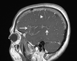


_driven_by_the_polymerization_of_actin_tails_(red).png)