Course:How Sex Differences Contribute to Anterior Cruciate Ligament Injuries
Knee Anatomy Overview
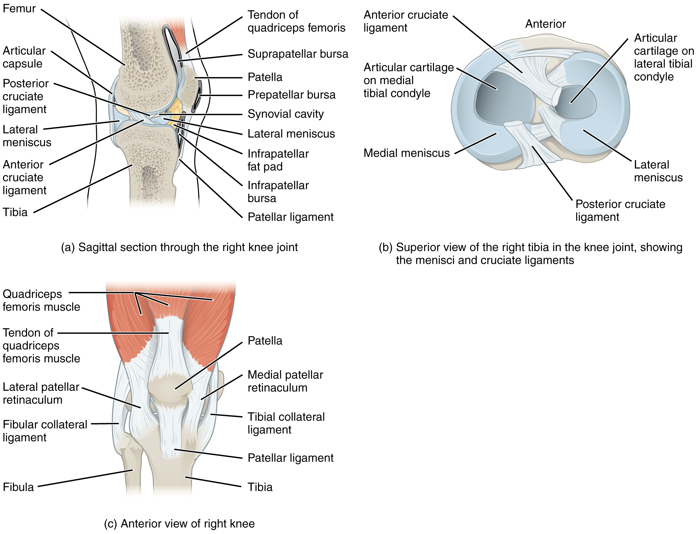
The knee is a synovial hinge joint composed of three articulating surfaces: between the femur and patella and medial and lateral condyles of the femur and tibia. There are four major ligaments in the knee[1]:
- Lateral Collateral Ligament (LCL): Prevents rotational and lateral-medial movement of the tibia with respect to the femur.
- Medial Collateral Ligament (MCL): Prevents rotational and lateral-medial movement of the tibia with respect to the femur.
- Anterior Cruciate Ligament (ACL): Prevents the femur from translating anteriorly with respect to the tibia.
- Posterior Cruciate Ligament (PCL): Prevents hyperextension of the tibia with respect to the femur.
Relative locations of each ligament are shown in this figure[1]. Note that fibular collateral ligament (FCL) and tibular collateral ligament (TCL) correspond to the LCL and MCL, respectively.
Some of the common injury pathologies for ACL injury are stiff landings (reduced knee flexion)[2][3], cutting maneuvers[4][5] and pivot landings (tibial rotation[6] and knee valgus landing[7]). The incidence rate of ACL injury in women may be as much as 10 times greater than in men [8].
Tissue properties of Male and Female ACL
The root cause of the injury discrepancy between males and females can be partially explained by looking directly at the tissues of the supportive structures. It has been found that there are substantive differences in anthropometry[9], ultra-structures[10] and material properties[9][11] between men’s and women’s ACLs; the former two driving the later.
Male ACLs are usually larger than female ACLs, most notably in terms of minimum area and total volume[9]. The magnitudes of these differences are exemplified below in Table 1:
Table 1: Male and Female ACL Anthropometric Data (reproduced from; Chandrashekar et al.[9])
| Males (n=8) | Females (n=9) | Percent difference (%) | |
| Length () | |||
| Minimum Area () | |||
| Volume () |
When analyzed on a microscopic level, the fibrous makeup of the ACL ligament can be examined. Hashemi et al. discovered that the number of collagen fibrils (the main material which ligaments are composed of) per unit area and the fibril area fraction (percent area of the sample with fibrils present) are substantially lower in female ACL samples [10]. These findings are presented below in Table 2:
Table 2: Ultrastructure Measurements of Human ACL (reproduced from; Hashemi et al.[10])
| Males (n=6) | Females (n=6) | Percent difference (%) | |
| Fibrils/Area (#) | |||
| Fibril Area Fraction (%) |
Together, the above differences culminate in substantively different material properties between the male and female ACL, as smaller material areas combined with diminished concentration of collagenous support structures reduce load bearing efficacy. Chandrashekar et al. found that female ACLs consistently produced lower values for load at failure, stress at failure and modulus of elasticity than male ACLs, suggesting that the female ACL may be less resistant to strain[9]. These findings are presented below in Table 3:
Table 3: Mechanical Properties of Male and Female ACL (reproduced from; Chandrashekar et al.[9])
| Males (n=8) | Females (n=9) | Percent difference (%) | |
| Elongation at Failure (mm) | |||
| Strain at Failure | |||
| Load at Failure (N) | |||
| Stress at Failure (MPa) | |||
| Stiffness (N/mm) | |||
| Modulus of Elasticity (MPA) |
Differences in Knee Injury Mechanisms Between Sexes
ACL injury was found to be common in sports involving jumping and cutting maneuvers such as basketball, soccer, and floorball, in which sudden decelerations are frequently performed[12]. For this particular type of motion, female athletes were found to be more prone to ACL injury compared to male athletes[4]. This may be due to females having an increased knee stiffness upon the instance of ground reaction force (GRF) impact compared to males[4], leading to higher load transfer to the knee joint. Increased knee stiffness may result in a smaller degree of knee flexion in women when decelerating[4][5]. The degree of landing stiffness is positively correlated with the GRF magnitude, leading to higher rate of anterior tibial translation and ultimately ACL injury.
Cutting
In a study by Miranda et al.[5] the jump-cut maneuver between the two sexes was studied. Subjects performed a cut at a 45 degree side-step onto a load cell which measured the magnitude of the GRF and used cameras to track the motion of each subject. The data showed that female subjects had a peak GRF of 1.45 times larger than the male subjects. Researchers were also able to examine the kinematics of the subjects to determine the knee flexion angle during physical exertion. They concluded that during contact between the leg and the ground, females exhibit less knee flexion angle excursion (knee joint flexion at the point of contact which acts to dampen the impact of the GRF)[5]. It is suspected that this may cause higher loads to be transferred to the knee joint, making it susceptible to ACL injury.
A separate study by James et al.[4] also found that females exhibited smaller maximum knee flexion (5.8 degrees less) when performing the cutting maneuver compared to males, caused by increased landing stiffness. By capturing strategically placed reflective spheres on anatomical landmarks with cameras, they were able to recreate the joint kinematics to calculate knee flexion angle using vector geometry. Furthermore, the GRF during the cutting maneuver was measured using a force plate. Findings by James et al. are illustrated in this figure[4] which shows GRF magnitude and knee flexion angle at ground contact for each male and female participant. The graph is separated into four quadrants, with the origin indicating the mean of the data points; quadrant IV represents the region of highest ACL injury risk potential corresponding to a high GRF and low knee flexion angle. It can be observed that since female test subjects are concentrated in quadrant IV, they may be more prone to ACL injury.
Jumping
In terms of jumping, Weinhandl et al.[3] studied GRF, kinetics and kinematics of males and females jumping from different absolute heights and a height equal to the max jump of each individual. Similar to that of cutting, the peak GRF of females were found to be significantly larger than males. As well, Leppanen et al. conducted a vertical drop jump (VDJ) test on 171 female athletes to explore if stiff landings are a factor in ACL injuries[2]. The study concluded that “decreased peak knee flexion angle and increased vertical GRF in a VDJ test were associated with an increased risk of ACL injury in young female team athletes”[2].
Pivot landing, where tibial torque is present during the landing portion of a jump, was found to be a factor in increased ACL injury risk for females as well. According to Beaulieu et al.[6], the worst loading scenario for the ACL is a combination of a tibial torque with a knee valgus moment. Russell et al.[7] also found that females tend to land in knee valgus while males tend to land in knee varus, therefore this injury mechanism may contribute to the increased ACL injury risk in women.
Conclusion
In conclusion, the studies above observe mechanisms of motion that propose a pathology for increased ACL injury risk in females compared to males. For both cutting and jumping motions, the two mechanisms that may be related to increased ACL injury rates are reduced knee flexion angle and higher GRF. Particularly in jumping, the tendency for females to land in the knee valgus position may also contribute to increased risk of ACL injury.
Strengths and Limitations in Literature
Tissue Properties of Male and Female ACL
A key strength in the work of both Chandrashekar et al. and Hashemi et al. is within their original hypothesis - there are substantive differences between male and female tissues[9][10]. This assumption is non-obvious and not the default starting point by others in the field, such as Woo et al., who simply endeavored to quantify the properties of ligaments and used samples from both sexes[11]. By starting with this base assumption, the authors were able to validate and quantify differences and highlight important findings instead of differences disappearing into an average value.
The major weakness in this work are sample size in some of the studies explored. There are large deviations in human samples due to many factors, such as age, activity level, diet etc., a fact noted by both authors, and this sample variation may lead to substantive variation in material property values. For example, the female modulus of elasticity presented in Table 3 has a window of values greater than the magnitude of the average value itself. Although with increased sample size the variability isn't necessarily reduced, having a greater number of data points to analyze would allow greater understanding and characterization of the apparent variation.
Differences in Knee Injury Mechanisms Between Sexes
The strength of the knee injury mechanism literature is the fact that they reach similar conclusions in regarding increased ACL injury risk for females. Each paper studies different motions (cutting and jumping) with methodological differences between authors, but tended to attribute injury to the same factors (increased knee stiffness, higher GFR)[3][4][5]. This supports and provides added confidence in the accuracy of conclusions.
However, with any study using video motion capture to track limb kinematics[3][4][5], there exist a number of limitations. Firstly, the fields of view may be restricted due to obstruction by other limbs and components. In cases where markers may be hidden from the camera, inaccurate vector calculations may result leading to false limb kinematics. Secondly, the motion tracking markers are prone to undesirable movement in high intensity maneuvers, such as the cutting and jumping motions described above. Thirdly, the placed markers are only an estimation of where each anatomical landmarks are located and may not accurately reflect true position during motion. These limitations were often noted by the authors of the referenced studies.
Controversy
Controversies in this field of research are minimal. Overall, the referenced authors were able to reach similar conclusions as to why the female ACL is more injury prone compared to the male ACL, whether the research was related to material testing or injury biomechanics of the ACL. However, a controversy in the studies that was apparent is in the difference between the likelihood of female ACL injury compared to males. As stated by multiple sources such as Toth et al., Cheung et al. and the Journal of Orthopaedics, the female ACL is either 2-8 [13][14] or 2-10[15] times more likely to be torn compared to the male ACL. This wide range in values suggests that consistency has not yet been achieved, and more research should be done to assess the likelihood of ACL injuries between sexes.
Another possible controversy is the limited sample sized used in material testing of the ACL. As seen in the above Tissue Properties section, referenced studies had sample sizes of 17 [9] and 12[10]. These small sample sizes may results not being representative of the population in question
Future Research Priorities
Although current research does suggest that females are more prone to ACL injury than males, some priorities for future work have also been identified.
Beaulieu et al.[6] produced a model to test the effects of fatigue failure in ACL injuries. Although results were not statistically significant, the group suggests that fatigue life may be a factor in higher rates of ACL failure in females, with smaller ACL cross sectional areas as a contributing factor[9]. Further experiments with a larger sample size are required.
James et al.[4] and Russell et al.[7] found that female subjects tended to land with a valgus knee alignment while male subjects tended to land with a varus knee alignment during cutting and jumping, respectively. The groups suggest that this difference may correlate with higher rates of ACL injury in females. Additionally, Beaulieu et al.[6] suggests that the most dangerous loading scenario for the ACL is a combination of a tibial torque with knee valgus moment. Further work is required to solidify the link between varus/valgus knee alignment and ACL injury over a range of loading scenarios.
Russell et al.[7] also proposes a correlation between varus/valgus knee alignment and muscle activation levels (males and females, single leg drop) but did not find statistically significant results. However, in a narrative review of existing literature Bencke et al.[16] reported that females may be at greater risk for ACL injury due to differences in quadriceps and hamstring activation, and closes by suggesting further research is required to explore the effects.
References
- ↑ 1.0 1.1 Open Stax (2019). 9.6 Anatomy of Selected Synovial Joints. In Anatomy and Physiology (Chapter 9). Retrieved from https://opentextbc.ca/anatomyandphysiology/chapter/9-6-anatomy-of-selected-synovial-joints/
- ↑ 2.0 2.1 2.2 Leppanen, M. (2016). "Stiff Landings Are Associated With Increased ACL Injury Risk in Young Female Basketball and Floorball Players". The American Journal of Sports Medicine. 45: 386–393 – via American Orthopaedic Society for Sports Medicine.
- ↑ 3.0 3.1 3.2 3.3 Weinhandl, J. (2015). "Sex differences in unilateral landing mechanics from absolute and relative heights". The Knee. 22: 298–303 – via ScienceDirect.
- ↑ 4.0 4.1 4.2 4.3 4.4 4.5 4.6 4.7 4.8 James, C. (2015). "Gender Differences among Sagittal Plane Knee Kinematic and Ground Reaction Force Characteristics during a Rapid Sprint and Cut Maneuver". Research Quarterly for Exercise and Sport. 75: 31–38 – via Taylor & Francis Online.
- ↑ 5.0 5.1 5.2 5.3 5.4 5.5 Miranda, D. (2013). "Knee Biomechanics During a Jump-Cut Maneuver: Effects of Gender & ACL Surgery". Medicine and Science in Sports and Exercise. 45: 942–951 – via Ovid.
- ↑ 6.0 6.1 6.2 6.3 Beaulieu, M. (2019). "Risk of Anterior Cruciate Ligament Fatigue Failure Is Increased by Limited Internal Femoral Rotation During In Vitro Repeated Pivot Landings". The American Journal of Sports Medicine. 43: 2233–2241 – via Sage.
- ↑ 7.0 7.1 7.2 7.3 Russell, K. (2006). "Sex Differences in Valgus Knee Angle During a Single-Leg Drop Jump". Journal of Athletic Training. 41: 166–171 – via U.S National Library of Medicine.
- ↑ Cheung, E. C. "Anatomic Factors that May Predispose Female Athletes to Anterior Cruciate Ligament Injury". Current Sports Medicine Reports. 14: 368–72.
- ↑ 9.0 9.1 9.2 9.3 9.4 9.5 9.6 9.7 9.8 Chandrashekar, N. (2006). "Sex-based differences in the tensile properties of the human anterior cruciate ligament". Journal of Biomechanics. 39: 2943–2950 – via ScienceDirect.
- ↑ 10.0 10.1 10.2 10.3 10.4 Hashemi, J. (2008). "The human anterior cruciate ligament: Sex differences in ultrastructure and correlation with biomechanical properties". Journal of Orthopaedic Research. 26: 945–950 – via Wiley Online.
- ↑ 11.0 11.1 Woo, S. (1991). "Tensile properties of the human femur-anterior cruciate ligament-tibia complex. The effects of specimen age and orientation". American Journal of Sports Medicine. 19: 217–225 – via Sage.
- ↑ Gianotti, S. (2009). "Incidence of anterior cruciate ligament injury and other knee ligament injuries: A national population-based study". Journal of Science and Medicine in Sport. 12: 622–627 – via ScienceDirect.
- ↑ Toth, A. (2001). "Anterior cruciate ligament injuries in the female athlete". Journal of Gender Specific Medicine. 4: 25–34.
- ↑ "The female ACL: Why is it more prone to injury?". Journal of Orthopaedics. 13: A1–A4. 2016 – via ScienceDirect.
- ↑ Cheung, E. (2015). "Anatomic Factors that May Predispose Female Athletes to Anterior Cruciate Ligament Injury". Current Sports Medicine Reports. 14: 368–372 – via Ovid.
- ↑ Bencke, J. (2018). "Muscle Activation During ACL Injury Risk Movements in Young Female Athletes: A Narrative Review". Frontiers in Physiology. 9: 445 – via Gale OneFile.